how do they x ray babies hips
The type of imaging used depends on your childs health and the reason why the imaging is needed. For example chest X-rays and hip X-rays require multiple images.

Legg Calve Perthes Disease Non Operative Treatment Wheeless Textbook Of Orthopaedics Disease Avascular Necrosis Pediatric Nursing
Ultrasounds are the diagnostic method of choice for infants under 6 months of age.

. The scan usually takes about 20 minutes. Because of the risk of developmental dysplasia of the hip in infants born breech-despite a normal physical exam-the American Academy of Pediatrics AAP guidelines recommend ultrasound US hip imaging at 6 weeks of age for breech females and optional imaging for breech males. During the examination an X-ray machine sends a beam of radiation through the pelvic bones and hip joints and an image is recorded on a computer or special film.
The hip ultrasound will show the healthcare provider the position and shape of the hip joint. The purpose of this study is to report US results and follow-up of. X rays CT scans and magnetic resonance imaging MRI scans may also be used.
We deliver faster than amazon. An X-ray does not show the bones in a young baby until at least 6 months of age and therefore a hip ultrasound is preferred. How do they x-ray babies uk.
How do they xray babies hips. Your baby will be placed on a table on their back or side. This device holds the joint in place while the babys skeleton grows and matures.
This line is used to measure the acetabular angle and as a reference for Perkin line. When should I order an X-ray rather than an ultrasound to diagnose a musculoskeletal problem in an infant. In babies with hip dysplasia the joint has not formed normally and the hips are prone to moving in and out of joint.
An X-ray is less useful in younger infants as the hips still contain a large proportion of. Around 6 months of age enough bone is present in an infant hip to make an X-ray more accurate than ultrasound. If a hip click is felt your healthcare provider will usually obtain a hip ultrasound to assess the hip joint.
A Hilgenreiner line connects the inferior tips of the iliac bones at the triradiate cartilage. Images gifs and videos featured seven times a day. Baby teeth x ray meme a new meme puts teeth on babies and the results are hilariou.
This image shows the soft tissues and the bones of the pelvis and hip joints. Youll be asked to partly undress your baby and take off their diaper for the test. Its use is to immobilize the child so that they don.
The most useful lines and angles that can be drawn in the pediatric pelvis assessing DDH are as follows. Infants may need assistance to keep still. Subsequent x-rays will track the hip joints progress.
Because they spin around the body taking multiple images CT scans can deliver radiation doses that are up to 200 times higher than an average chest X. Imaging helps the orthopedist determine the deformitys location magnitude and. You will go in the room with him he will need to be stripped from the waist down they will take x-rays of him flat on his back legs dead straight and together you wil be able to hold him in this position then an x-ray of his still on his back with his knees bent facing outwards and the soles of his feet put together he will be fine its not traumatic at all you will.
Two tests are performed called the Barlow and Ortolani tests to examine the function of the hip joints. Wednesdays tragedy is thought to be the single biggest loss of life during the current migrant. Usually both hips are scanned.
How they x ray babies meme. Treatment for newborns A baby born with a dislocated hip can be successfully treated with a Pavlik harness. Pediatricians do often check for hip problems in babies and hip dysplasia is the most common hip developmental deformity in children.
Each hip should be evaluated from the side and front view and many times images of the hip in a flexed or extended view are needed to assess if the hip joint is. This image shows the soft tissues and. Up to 10 cash back Dog X-rays usually start around 200 and increase from there depending on how many images are needed.
A hip click can be felt by the examiner when the hip joints may not have formed normally. Typically a doctor will get the patient history do a physical examination and order a standing-alignment X-ray or EOS imaging of the leg bones from the hip to the ankle. It is estimated by the Center for Disease Control CDC that 1-2 of every 1000 babies have hip dysplasia.
During the examination an X-ray machine sends a beam of radiation through the pelvic bones and hip joints and an image is recorded on a computer or special film. An X-ray may be used in older children or adults to demonstrate the underdeveloped socket of the hip. If you are referred for an investigation or treatment involving ionising radiation you should inform clinical staff that you are or may be pregnant so that they can advise you.
The X-ray image is black and white. However many more go undiagnosed as it may be too mild to even detect. How do they X-ray babies hips.
The doctor hears or feels a hip click when moving the infants thigh outward during a routine checkup. The babys legs have differences in their lengths or appearances.

Hip Joint Developmental Hip Dysplasia 1 Year Old Child With A Dislocated Right Hip The Degree O Developmental Dysplasia Of The Hip Radiography Subluxation

X Rays Of Normal Hip And Hip With Arthritis Hip Replacement Total Hip Replacement Medical Laboratory Science

Lower Limb Radiographs Anatomy Anatomy And Physiology X Ray

Diagnosis Prevention And Management Of Canine Hip Dysplasia A Revie Vmrr Canine Hip Dysplasia Diagnostic Imaging Total Hip Replacement

Severe Hip Dysplasia In A Boxer The Red Arrows Are Pointing To The Over Growth Of Bone At The Femoral Neck Head T Shades Of Grey Hip Dysplasia Animal Heads

Causes Of Ddh Hip Dysplasia Baby Developmental Dysplasia Of The Hip Baby Wearing
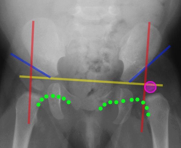
Lines Of The Hip Pediatrics Pediatrics Pediatric Nurse Practitioner Pediatric Radiology
How To Position The False Profile View X Ray Of The Hip X Ray Profile View Positivity

Pin On Hips

Pin On X Rays

Pin On Pavlik Harness

Pin On Fibro Autoimmune Diseases

Canine Ofa Hip Xrays Goldendoodle Puppy For Sale Labradoodle Goldendoodle Goldendoodle Puppy

Uk Professor Says Swaddling Epidemic Gives Babies Clicky Hips Daily Mail Online Hips Professor Baby Swaddle

Hip X Ray Loss Of Shenton S Line Is A Sign Of Fx Neck Of Femur Radiology Medical Technology Radiology Student

How To Shower After Hip Replacement Surgery Livestrong Com Hip Replacement Surgery Hip Replacement Exercises Hip Brace

Basic Information About Dog Hip Dysplasia Paperblog Dog Hip Dysplasia Hip Dysplasia Canine Hip Dysplasia

Developmental Dysplasia Of The Hips Start Screening For This At Age 2 Weeks With Barlow Nursing School Tips Developmental Dysplasia Of The Hip Nursery Nurse

Pin On Ortho